
Anatomy of the Heart
The heart is a complex and fascinating organ. It starts beating around 3 weeks after conception in the womb and continues to pump without fail during our lifetime (1). The heart beats about 100,000 times each day and pumps 2,000 gallons of blood throughout the body (2)!
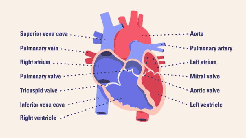
The heart is mainly made up of muscle called myocardium and comprised of four chambers or “rooms.” The two top chambers are the right atrium and left atrium. The two bottom chambers are the right ventricle and left ventricle. There are also four valves in the heart located after each chamber that act like doors and help control the blood flow through the heart. The valves are called the tricuspid, pulmonic, mitral and aortic valves.
De-oxygenated blood flows into the right atrium from the two large veins in the body called the superior vena cava and inferior vena cava. The blood then travels through the tricuspid valve into the right ventricle, though the pulmonic valve and into the pulmonary arteries that go to the lungs. The blood receives oxygen from the lungs. The oxygenated blood flows back to the heart via the pulmonary veins, into the left atrium, through the mitral valve and into the left ventricle, then through the aortic valve and into the aorta, which is the largest blood vessel in the body. The blood will then travel throughout our body to our other vital organs. For more information of the valves of the heart please refer to the “Valvular heart disease” section on the website.
When the heart “pumps,” this refers to when the ventricles contract. The left ventricle pumps the blood out of the heart, and the pressure that allows the blood to flow in the blood vessels in our body is called the systolic blood pressure (or top number on a blood pressure reading). The bottom number or diastolic blood pressure which is the pressure in our blood vessels in our body when the left ventricle is relaxed and filling up with blood.
The coronary blood vessels or arteries are the blood vessels that supply the heart organ itself. There is a right and left coronary artery. The left coronary artery splits into two branches – these are called the left anterior descending artery (that supplies the front of the heart) and the left circumflex artery (that supplies the left side and part of the back of the heart). The right coronary artery supplies the right side and back of the heart. For more information on the coronary arteries, please refer to the “Coronary artery disease” section on the website.
References:
1) Biga, Lindsay M, et al. (2019, September 26). Development of the Heart. Anatomy & Physiology. OpenStax/Oregon State University. https://open.oregonstate.education/aandp/chapter/19-5-development-of-the-heart/
2) Anatomy of a Human Heart (2019, February 27). Michigan Medicine. https://www.michiganmedicine.org/health-lab/anatomy-human-heart
3) Heart video: https://www.michiganmedicine.org/health-lab/anatomy-human-heart